
Scientists are learning more and more about the brain every year that passes. Will it have any effect on the way teachers teach and students learn? This fascinating question was brought alive when two discussion groups got together one evening to quiz a cognitive neuroscientist working on exactly this topic.
Educational practice has traditionally developed on the basis of theories – scientific or anecdotal – about behaviour. Psychologists study motivation and reward, thinking and remembering; sociologists look at family, peer groups and social class, while economists measure the effects of socio-economic background and the incentives of the jobs market. Cognitive neuroscience, the new kid on the block, is now adding to the mix.
The discussion group was keen to find out first, just what a cognitive neuroscientist is. Their guest had originally trained as a biologist, and she later became interested in cognition, the study of how we think. Functional magnetic resonance imaging (fMRI) was the tool she mainly used to study the brain, more specifically, the area of the brain activated when we think about people and things: the prefrontal cortex.
Functional MRI
MRI scanning is becoming widely known as a routine diagnostic tool for getting a clear, high resolution image of the interior of the body. It’s based on an amazing laboratory discovery, only a few decades ago, that atoms can be made to give off a signal when they are put into a strong magnetic field and then subjected to a rapidly oscillating radio wave. Although this was essentially a discovery in pure physics, it’s value for medicine was soon realised. The key point is that hydrogen atoms, abundant in almost all the molecules in the body, can be made to emit tiny amounts of energy when the scanner’s magnetic fields are switched on. The location of all these atoms is picked up by the machine. Specific chemicals can be added to increase the contrast between different types of tissue. With the development of computer technology the quality of images has developed to such an extent that features measured in millimetres can be detected.
The word ‘functional’ is used as prefix to MRI to indicate when the technology is being used to study a particular function rather than to simply locate atoms. A particular use was discovered in relation to the brain when it was found that the scanner can detect changes in the flow of blood to specific areas of the organ. With advances in processing the tiny signals from the blood vessels in the brain, it is now possible to locate quite precise areas of the brain associated with particular functions. When the nerve cells (or neurons) in these areas are active, blood flows preferentially to them. The resulting signal is processed by a computer which maps the location in relation to the whole brain. Examples of fMRI scans are shown below. The red indicates areas of the brain activated while the subject is using their working memory.
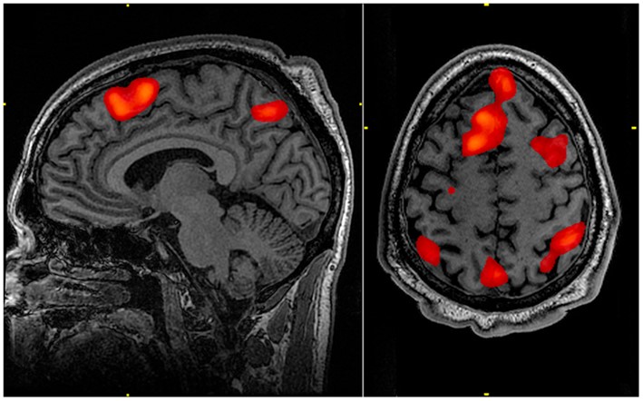
fMRI image of areas of the brain highlighted during a specific mental task
Over the long history of brain research various areas of the brain have become associated with particular aspects of our behaviour. Some, for example, have been shown to be linked to language processing, others to inhibiting emotions and another to processing visual information from the eye. Historically much of this evidence came from particular injuries that gave rise to specific behavioural changes, such as loss of inhibition or impaired language processing. The diagram indicates some of the major areas of the brain.
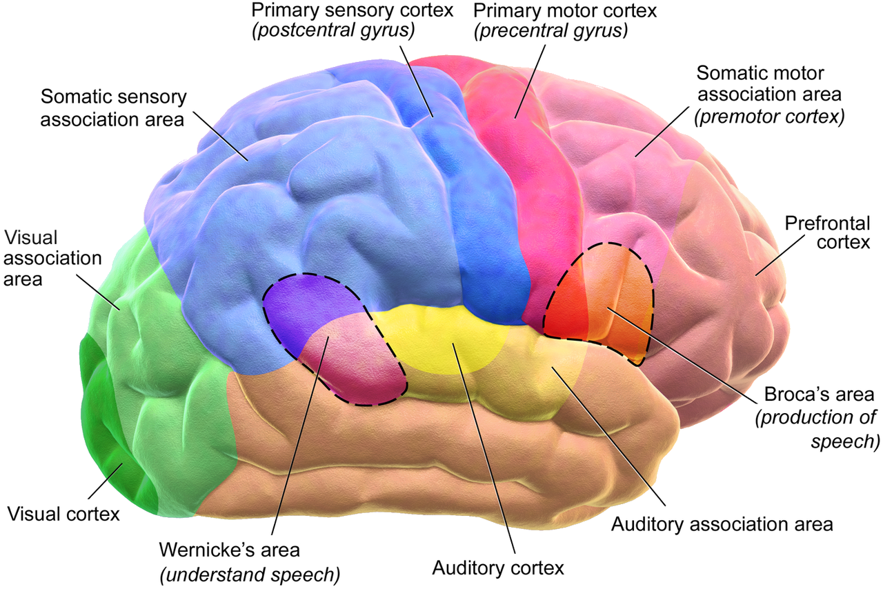
Areas of the brain
(Courtesy of Medical gallery of Blausen Medical 2014 1)
Neurons and synapses
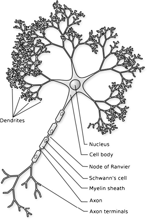
The discussion group was keen to find out about the way the brain develops from the infant to the adult stage. Surprisingly, it turns out that at birth you have the maximum number of neurons you’re ever going to have. A neuron is simply a long, thin nerve cell which conveys an electrical signal from one of its ends to the other. Each neuron is connected to a number of other neurons (maybe thousands) at a junction called a synapse. The number of neurons at birth is about 1000 billion. If each of these may be connected to up to thousand others, you get an idea of the extraordinary scope for complexity offered by the network of neurons in the brain.
Diagram of a neuron
At birth however, despite having a full complement of neurons, the brain has relatively few connections. As the baby grows, more and more ‘branches’ get made in networks in a process with the delightful, tree-like name of ‘arborisation’. These form a growing number of connections (or synapses) with other neurons. The density of synapses continues rising until it peaks around the age of 2 -3 and thereafter it plateaus.
An even more important discovery is that those connections that get used most also get maintained, whereas underused ones disappear. This remarkable phenomenon is captured in the phrase: “neurons that fire together, wire together”.
This discovery seems to reinforce many commonly held ideas and practices, for example that “practice makes perfect” in sport, music and language learning or that being mentally active in old age – reading, crosswords, puzzles, quizzes – helps stave off dementia.
This fascinating process, in which some connections (or synapses) disappear and others are strengthened, goes by the equally arboreal name of ‘pruning’. In infants each neuron makes an average of 2,500 synaptic connections and this peaks at about 2-3 years of age to some 15,000. Thereafter the neuron connections that are used least gradually disappear. This pruning” process is dependent on environmental factors and external stimuli. It is, in effect a process of learning.
One person in the group asked why neuron connections are made in the first place if they are then pruned away? It appears that, because every connection demands a certain amount of energy, it’s efficient to only sustain those that are needed. Another wondered whether some connections might be pruned away that are needed later. Apparently much depends on the age at which the pruning happens. In a study of kittens reared with only vertical lines in their visual environment they are unable to see horizontals after a certain age.
Learning
The act of learning enhances the development of connections between neurons. A study of juggling for example shows that learning to juggle leads to an increase in the density of synapses. After the initial learning this settles down to previous levels even though the skill remains. There is a temporary increase during learning. This seems to correspond closely to the experience of acquiring a skill, whether playing the piano, throwing the javelin or driving a car. At first considerable conscious effort is involved, but, with repeated practice, the action become increasingly automatic. Our neuroscientist pointed out that as the so-called “muscle memory” develops – enabling pianists and typists alike to perform automatically – activity in the brain moves from its initial place in the pre-frontal cortex to the posterior parts. Interestingly she added that studies have shown that children who learn to skip first also tend to learn to read first.
With new insights brought by the development of fMRI and other technologies, people interested in education have lost no time in trying to work out how the new knowledge might affect the way we teach as well as learn. One example is in the area of memory. How frequently should you repeat an act of memorising, what kind of delay is best before you practice retrieving it? This was an area our neuroscientist was herself working on. It turns out you remember better if you repeat after a matter of days. A number of different factors influence our ability to remember things, including motivation and attention. I expect these scientific insights only go to confirm what we know all too well from our own experience of trying to recall things in different moods.
Rewards and risks
Many practices in education are, as you may well recall, based on rewards. Were you ever offered brownie points for behaving well? Trials are underway to study the effects of various kinds of reward on improving the ability to control behaviour and concentrate. The discussion moved on to gratification – one person asked whether deferred and immediate gratification were linked to the same part of the brain. It turns out there are important differences. Deferring requires you to think before you act and that brings in the thinking area of the brain: the frontal cortex.
Risk-taking is another area that seems to involve changes as we grow up. Young males seem particularly prone to excessive risk-taking. Our neuroscientist was working with a colleague on an experiment on this very topic. They had set up a game that simulated a risky road-crossing and invited people of different ages to play, giving incentives to take risks. The number of ‘accidents’ that followed was recorded. Although initially there was little difference in risk-taking between the adolescents and adults, when friends were brought in to accompany the risk-takers the adolescents’ scores doubled. The implication seems to be that the social context acts as a reward, encouraging risky behaviour. Again, a behavioural trait that seems to chime with anecdotal experience of group behaviour. It seems there may be evolutionary reasons for this aspect of teenagers’ brain function. Given that our physiology is largely adapted to the environment of early Homo sapiens it’s likely that in the teenage period those individuals who took greater risks were more likely to find a mate and create a place to bring up offspring safely. That’s how evolution works – those that fit the environment best tend to survive longer and have more offspring. Their genes are therefore more likely to be present in subsequent generations.
Education
Cognitive neuroscience lends itself to application to real-world problems – in education, for example. It has been discovered recently that when a child (or anyone else) moves on from a more primitive to a more advanced concept, the naive ideas they had beforehand (like the Earth being flat) are not necessarily expunged. Instead they hold on to them alongside the new scientifically-based knowledge. If the new concept conflicts with the evidence they believe (e.g. the Earth being visibly flat), a particular area of the brain (the cingulated cortex) detects the conflict. The process of learning involves inhibiting, rather than eliminating naive ideas. Our neuroscientist was working with teachers to develop a tool to help children inhibit their naive interpretations of things, opening up the way to more advanced understanding.
This insight into the multi-disciplinary world, in which technologists, biologists and teachers collaborate to gain insight into the learning process, shows something of the diverse ways in which science and technology can be applied – when in the right hands.
© Andrew Morris 16th May 2019
