
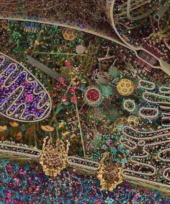
This alluring image caught the attention of a science discussion group recently. Looking at first sight like a rich middle-Eastern tapestry, it is in fact a model of part of a typical human cell put together using information from various instruments used to study tiny things.
Its aesthetic appeal drew the group members in and the complexity of its composition provoked questions about what it represented.
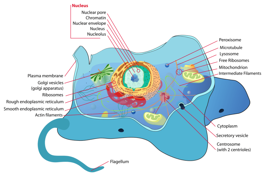
Conventionally, images of the cell in books and websites emphasise the variety of parts and their technical names – as the diagram on the right demonstrates admirably. It looks daunting – a bit too much to take in, unless you’re preparing for an exam.
Curiosity amongst the discussion group was not so much about the parts, but the thing as a whole. How big are cells? How many have we got in our bodies? What do they do? How do the bits interact? Are they busily working away all the time?
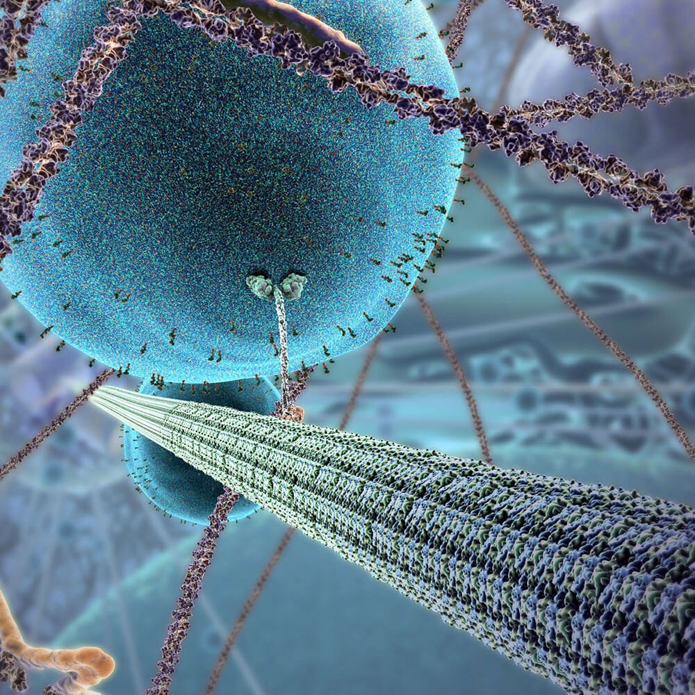
Pictorial representations of a cell – or any other biological item – are traditionally static and two-dimensional. In reality, however, cells are buzzing with activity, as are the molecules of which they are composed and the tissues to which they contribute. Advances in animation technology and biological imaging have now brought this activity alive for us – in glorious 3-D technicolour.

This 3-minute example from Harvard University The Inner Life of the Cell shows molecular structures interacting inside a typical cell. Although it doesn’t explain what’s happening in detail, it does give an impression of the shape of structures and the way in which they interact with one another. This particular clip follows a white blood cell’s movement along the lining of a body tissue and its response to an external stimulus.
Scale
The first issue for the discussion group to sort out was the relative scale of things. Molecules, cells, viruses, bacteria, DNA …… which is smaller than which?
Let’s start with the cell – it’s the main unit from which animals and plants are built. Individually, cells are created, then develop, reproduce and die; together they form the tissues of which organs are made (though there are some exceptions to each of these points). Cells come in numerous types which vary enormously in detail; but an important common feature of all cells is the outer membrane, separating the inside from the outside. The watery world inside a cell and the watery world outside are separated by an oily boundary that protects the workings of the interior. Like prison cells, what goes on inside is quite unlike what happens outside. The boundary is closed to all but carefully selected molecules …. until a virus finds a way to trick its way in, that is. That’s what the coronavirus has achieved.
Cells are relatively large items, at the microscopic level – typically measured in micrometres (thousandths of a millimetre), though sizes do vary considerably. The structures inside a cell are typically ten to a hundred times smaller. Many of these structures are built out of giant molecules, often proteins, which might be ten to a hundred times smaller still. The coronavirus is made of giant molecules and measures about a tenth of a micrometre – perhaps a hundred times smaller than a typical cell. This diagram gives an impression of relative sizes. Note that the steps in the horizontal direction are not equal – each one is ten times larger than the previous one.
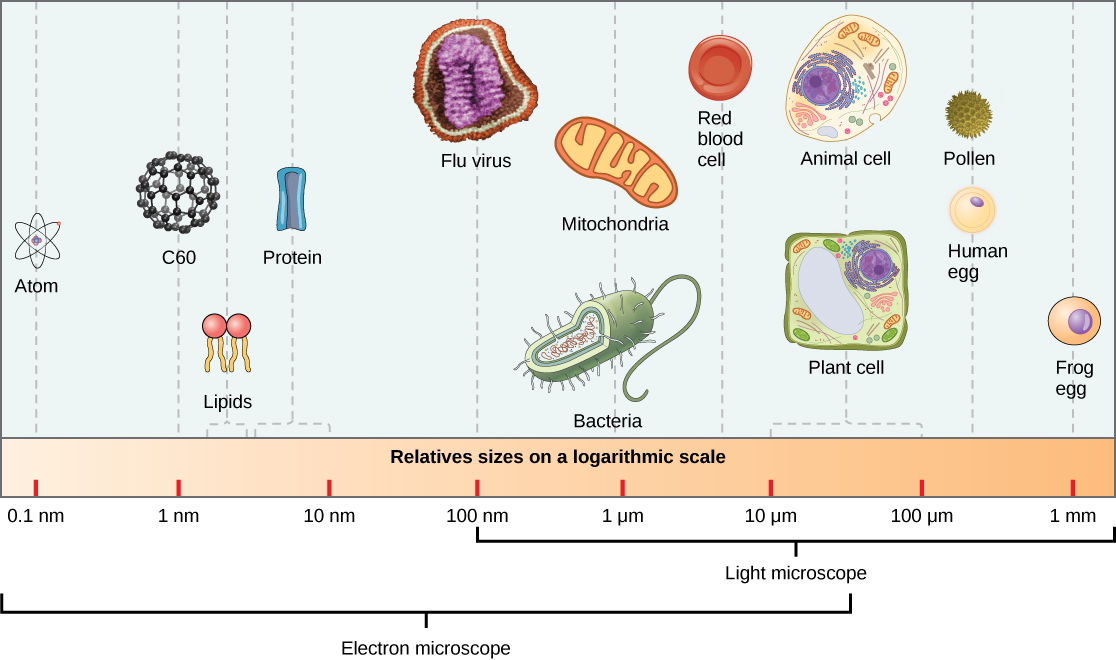
(from Concepts of Biology courtesy of Charles Molnar and Jane Gair)
Tissues and cells
Cells aggregate together to form tissues of various type – connective tissue or muscle tissue, for example. Organs, such as kidneys or lungs are made up of various tissues. In the heart, for example, you find muscle tissue, connective tissue and nerve tissue. Cells aggregate to form tissue and tissues to form organs.

To survive, an organism needs to carry out a huge range of functions. To serve all of these, cells are of many types, each adapted for its specialised function. Nerve cells transmit electrical pulses for example, muscles cells contract and red blood cells carry oxygen around.
On the left are images of a red blood cell, a platelet and a white blood cell (looking a bit blue), taken with an electron microscope.
blood cells
Below, on the right are muscle cells. On the left, in bright green, a nerve cell (neuron) from the brain.
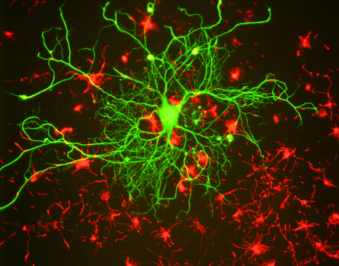
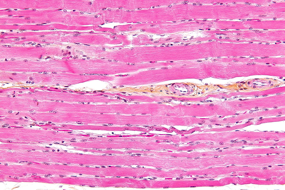
As these images demonstrate, the shapes of different kinds of cell varies enormously. Their forms may even express something of their function: the voluminous red blood cell stores vast numbers of haemoglobin molecules; the wiry looking neuron transmits electric pulses; the long, thin muscle cell elongates and contracts.
Cells are, of course, mostly tiny – powerful microscopes were needed to create the images above. They vary in size enormously: red blood cells being typically a few micrometres (thousandths of a millimetre) across, whereas some nerve cells may extend for metres. The enormity of the number of cells of which we are made is as difficult to grasp as the tiny size of the average cell. It’s reckoned there may be 30 to 40 trillion in a human being, and that’s without counting the bacteria.
Inside the cell
The first two images above give an impression of the internal components of a cell (known as organelles – small organs). The animation mentioned above, The Inner Life of the Cell, gives some idea of the activity that goes on inside these structures. Tiny as most cells are, they are complex environments, filled with smaller units, each with a vital role in maintaining the life of the cell. They are more like a walled city than a village.
Essential to life is the cell’s ability to take in and use energy. The components inside cells responsible for this are discrete bodies with their own outer membrane, called mitochondria. Remarkably, all organisms, not just animals, make use of the same energy-rich fuel: glucose. It’s a simple molecule, a kind of sugar, that the body obtains by breaking down the molecules in the food we eat. It travels to all the cells of the body via the blood – that’s why a blood test can reveal the level of glucose in your body.
There are many mitochondria in a typical cell (maybe thousands). Inside each is a set of enzymes which combine the energy-rich glucose from food with oxygen from the lungs. Rather like burning oil or coal in air, this releases energy, which is immediately stored. A high-energy molecule common to all forms of life (known as ATP) takes up and stores energy ready to be used wherever and whenever needed.
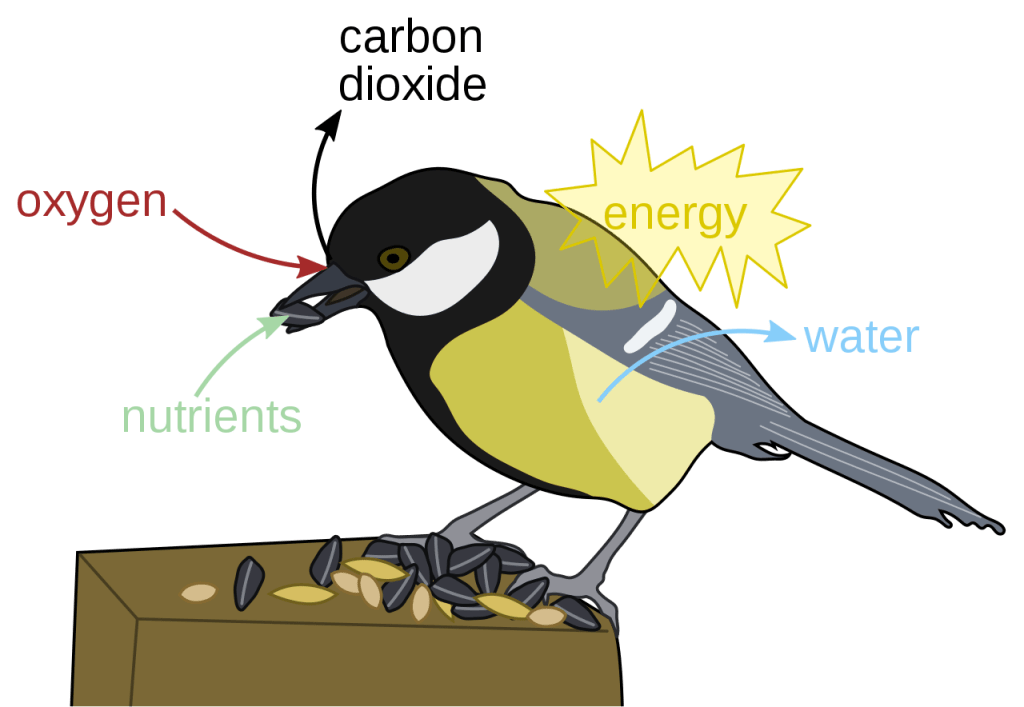
This is why we need to breathe in oxygen – every cell requires it all the time to release energy, just as a motor vehicle does to burn petrol. As matter of interest, the waste products of this process (called respiration) are carbon dioxide and water – that’s why we end up expelling these particular substances
Respiration process (image by Nefronus)
Another vital function of the cell is to store, and ultimately reproduce, the vital genetic information that it will need to pass on when it divides into daughter cells. This information resides in the enormously long DNA molecules, held in another organelle: the nucleus. Most cells have just one of these. Like mitochondria it is also separated from the rest of the cell by a membrane.
In everyday life we come across DNA in connection with our identity and heredity: identifying individuals from DNA samples or observing genetic similarities in family members. But DNA molecules are not just waiting around for the occasional moment in which they get passed on to the next generation. Their day-to-day work is to provide the information need to make proteins. Genes are short stretches of the long DNA molecule. Each one carries information specifying the make-up of a particular type of protein. One gene carries the code for a digestive enzyme, another the code for a muscle protein, another for a particular type of antibody. Altogether, there are tens of thousands of types of protein in the body and there’s a unique gene for each one. Information coded in the DNA molecules stored in the nucleus of a cell, is being used continuously to produce the proteins needed to maintain life.
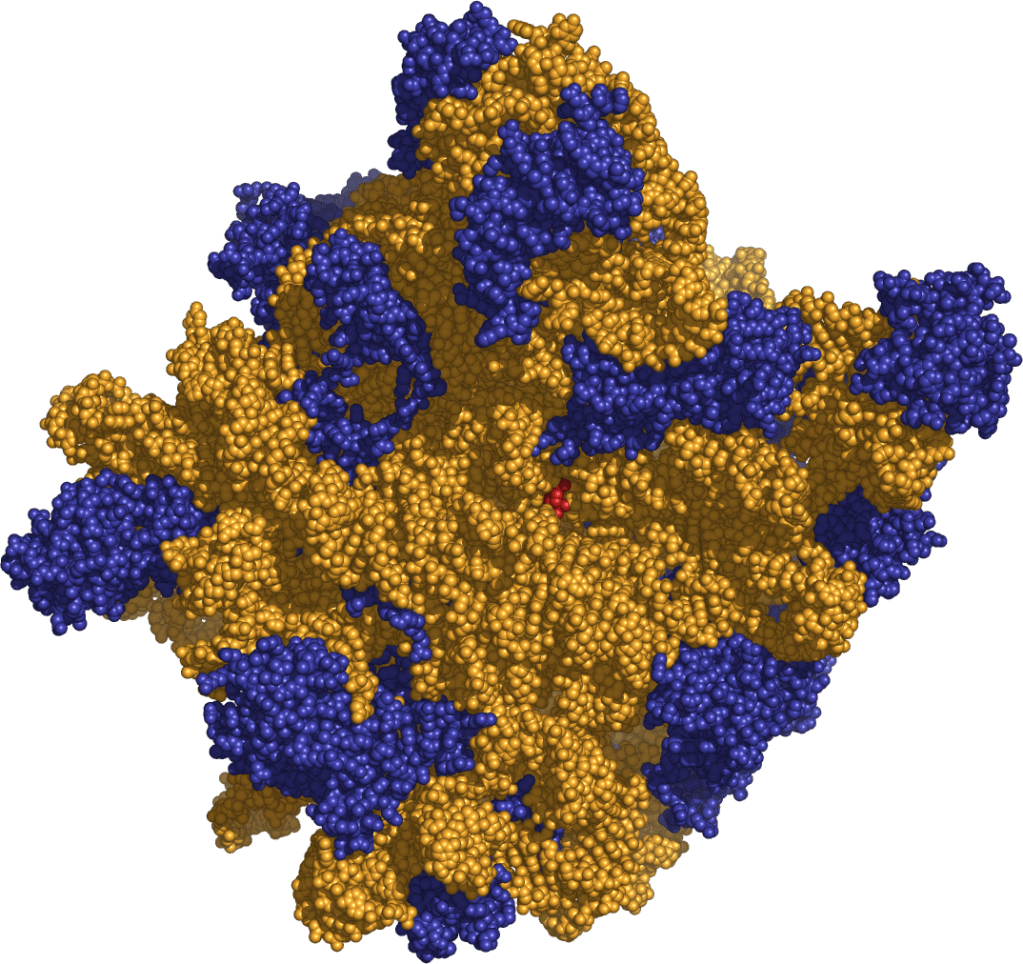
A third type of organelle is the site at which proteins are produced, using information encoded in the genes. Known as ribosomes, these float around in the gel-like environment inside a cell (known as protoplasm). A cell in our body may contain literally millions of these.
As this model shows, large assemblies of molecules, such as ribosomes, may be made of hundreds of thousands of atoms (the orange and blue balls).
Ribosome: a model of one subunit
It is clear that the inside of cell is a complex and busy place! We’ve introduced just three types of organelle: the mitochondrion, nucleus and ribosome; several more remain to be explored. What struck members of the discussion group at this point however was not the desire for further detail about the complexity, but rather, the need to pause and reflect on the almost unbelievable facts already presented. There are trillions of cells in our bodies, each of which contains millions of “moving parts”. Powerful microscopes are needed to visualise cells and even more sophisticated equipment – electron microscopes and X-ray diffractometers – to do the same for the molecular structures that carry out activity within the cell. It’s difficult to take in the miniscule size of the structures that make our bodies work and the enormous number of them in each of us.
Molecules at work
The multitudinous structures that make up our cells are mostly assemblies of giant molecules, such as proteins and DNA.
The animation mentioned above (The Inner Life of the Cell), portrays large protein assemblies moving in and around a cell. Although it doesn’t explain what exactly is going on, it gives a vivid impression of some of the actions of which proteins are capable. In one sequence a long, thin molecule is shown as having two “legs” each capable of shifting between two positions. Alternating movement between them gives the impression of ‘walking’ along a molecular fibre.
In a later sequence, one protein (an enzyme) cleaves a long thin molecule in two – breaking bonds between atoms in this way is what a whole class of enzymes do. It’s how the giant molecules in your food get broken down into simpler ones.
A topical activity in the current pandemic is the way antibodies act against viruses. In this animation, antibodies neutralise a flu virus. It shows the structure of a whole virus, its entry through the membrane into a cell and the entry of the virus’s RNA (like DNA) into the cell’s nucleus. It shows how antibody molecules attach to a newly replicated virus, after it has been made, in such a way that the new virus is unable to get back in to the cell’s nucleus to replicate itself. Unable to replicate and spread to other cells the virus is effectively neutralised. The equivalent mechanisms for coronavirus remain to be discovered.
What to make of it all
The question that arose, as the group contemplated the intricacy of the activity within a cell, was just how rapidly it all happens – is it incessant, does a cell ever rest? There’s no simple answer to the question of pace. The enzymes which break bonds and alter the shape of molecules are acting as catalysts. This means they dramatically speed up the rate of chemical processes in the body, compared to the corresponding speed in a laboratory experiment. Some enzymes perform 100s of reactions in a second, others only one a minute. For comparison, an average typist manages just over three characters in a second – maybe a hundred times slower than an enzyme’s action. On the other hand, an estimate of the time taken for DNA to replicate itself when a cell divides, is of the order of 10 hours. The take-away message seems to be that chemical processes are happening in unimaginably great numbers all the time in the body, but the time taken for them to happen varies from a tiny fraction of a second to a sizeable fraction of a whole day.
The demands on a cell will vary depending on its type and on the circumstances of the moment: digesting, sleeping, running, thinking. Simply maintaining itself is a ceaseless activity for a living cell. There is never complete rest from biochemical activity in the cells of the body. No wonder we need a good long sleep at the end of the day – and even while we lie dreaming we’re still pretty active, underneath the skin.
© Andrew Morris 27th November 2020
To be alerted when a new blog is published email andrewmorris110@gmail.com
